I
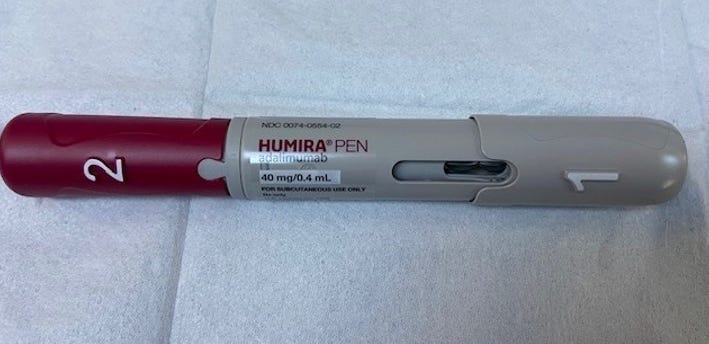
was given this medication by a patient with Crohn’s disease for
analysis. It is used to treat various inflammatory conditions, including
rheumatoid arthritis, psoriatic arthritis, ankylosing spondylitis,
Crohn's disease, ulcerative colitis, plaque psoriasis.
I had previously shown microelectronics in Enbrel:
Darkfield
Microscopy Of Pfizer's Enbrel (Etanercept) Shows Nanoantennas,
Microrobots, Self Assembly Nanotechnology Comparable To C19 Bioweapons.
Microelectronics For Patient Monitoring
What Is Enbrel Injection Doing To The Blood? Darkfield Live Blood Documentation
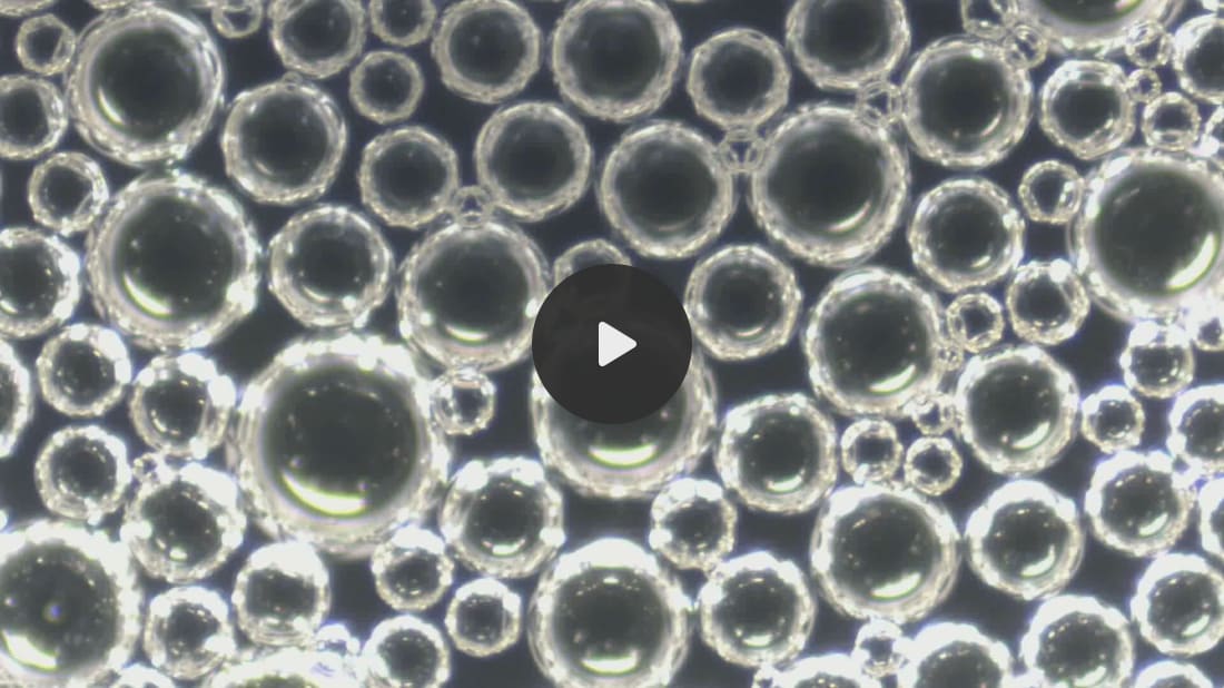
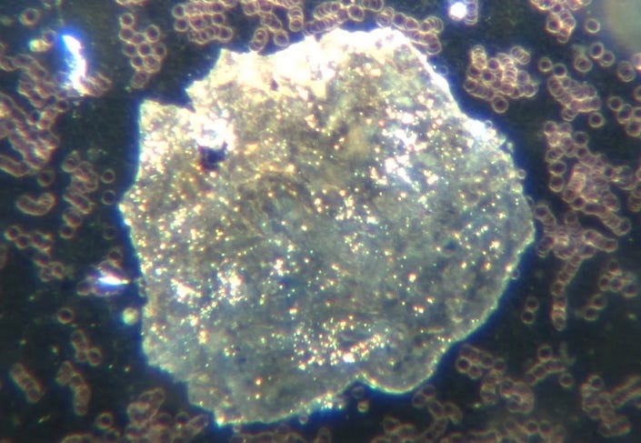
These are liposomes in this medication seen at 100x magnification:
I showed similar liposome images in my analysis of Pfizer COVID19 bioweapon.
Pfizer
BioNTech COVID19 Bioweapon: Self Assembled Liposomes After 2 Weeks At
Room Temperature. Correlations To COVID19 Vaccinated Embalmed Blood And
Unvaccinated Live Blood - Its All The Same!
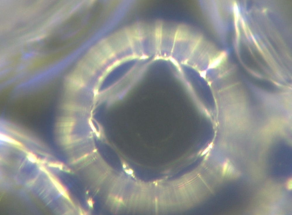
Here is an image of its internal geometry - magnification 2000x.
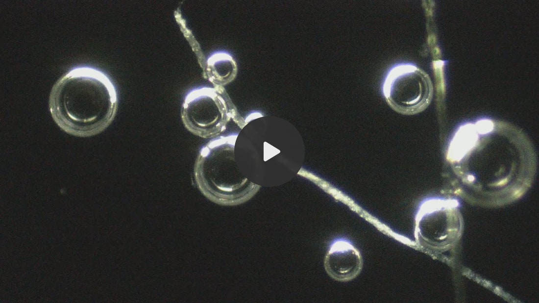
Here is interaction with polymer filaments - magnification 200x. :
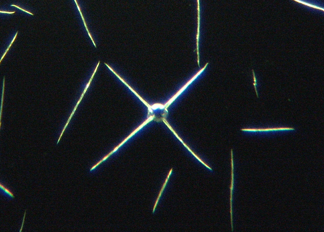
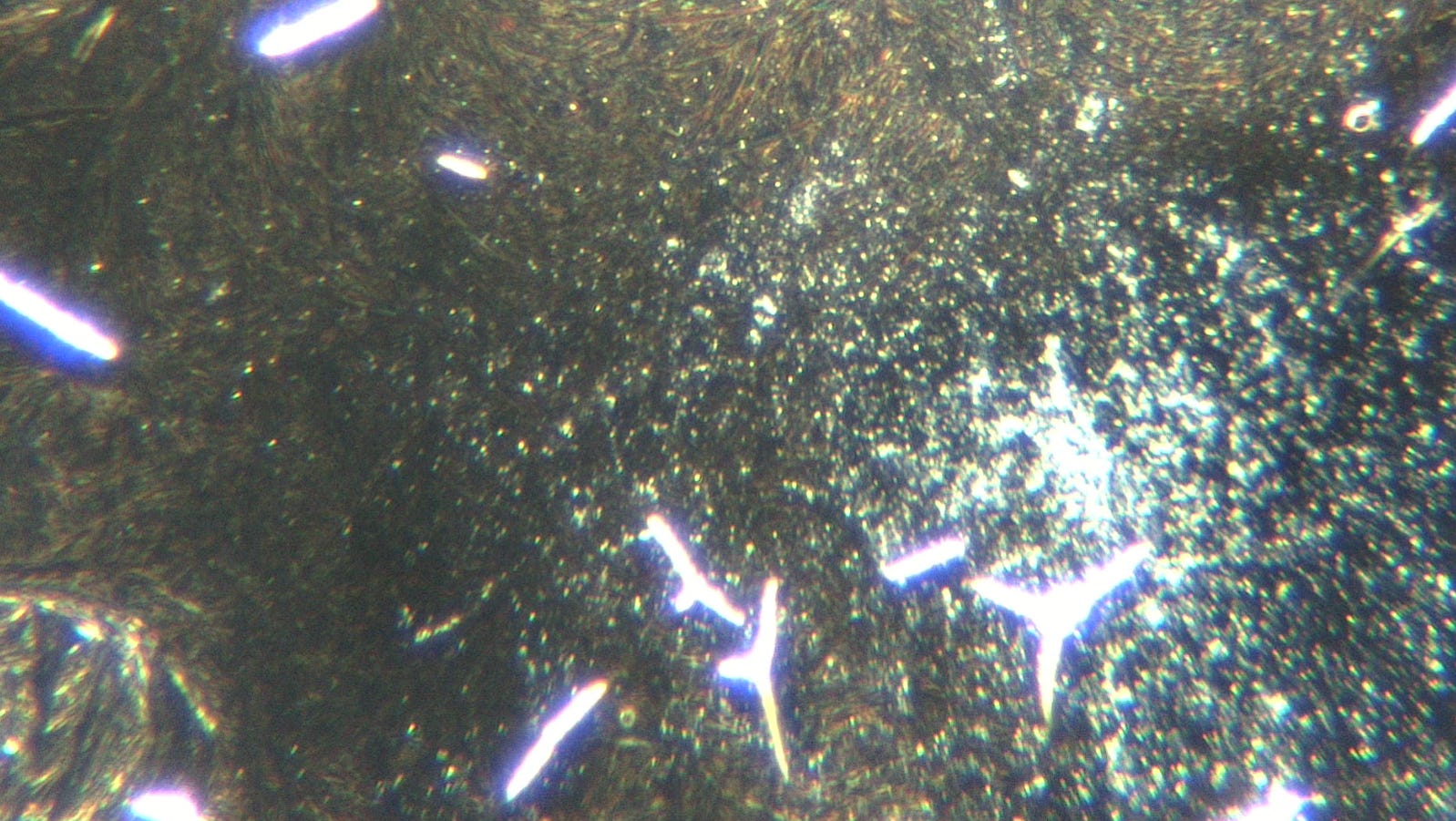
The liposomes developed antennas - magnification 200x.
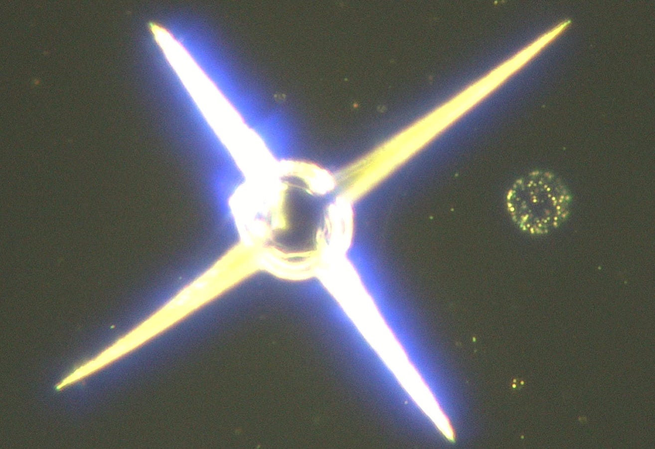
Luminescence seen with adjacent microrobot - magnification 2000x.
Here the liposomes and their arms connect and change luminescence to dark - magnification 200x.
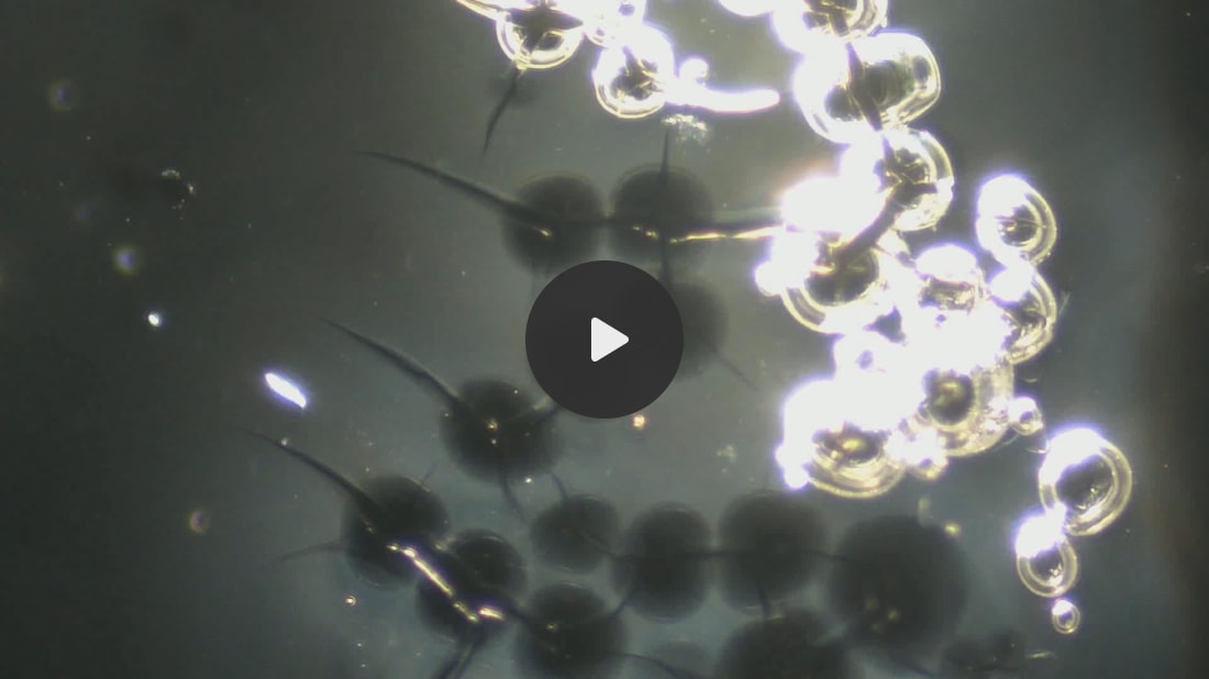
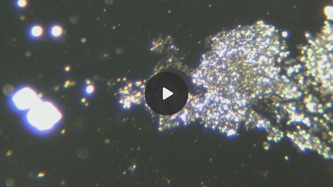
Here accumulation of microrobots into mesogens is seen - magnification 200x.
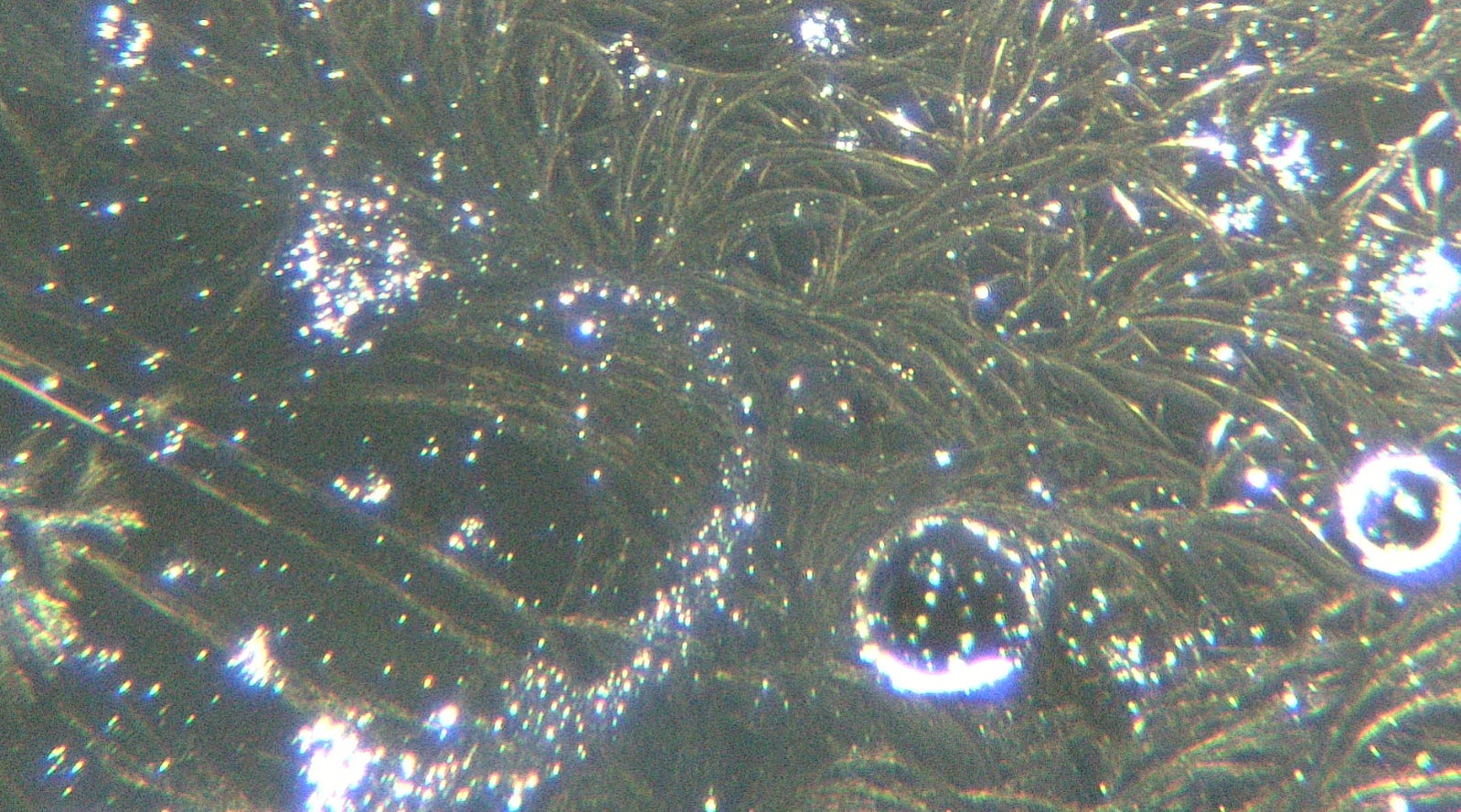
The
liposomes spin nanofibers into elaborate networks interspursed with
microrobots - magnification 200x. Liposomes are seen as the origins of
this network:
The liposomes also attract microrobots as well as surrounding nanofibers - magnification 100x.
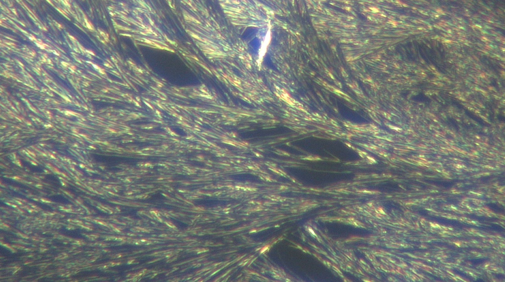
Here the elaborate nano/ microfibers are seen - magnification 2000x.
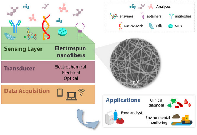
Nanofibers are used for biosensing applications:
Nanofibers interfaces for biosensing: Design and applications
The
composition of NFs can also be modulated to increase the number of
sites for immobilization of recognition elements, as well as to enhance
the interaction with the target analytes [44].
In this regard, a plethora of synthetic and natural macromolecules, as
well as their blends, have been processed by electrospinning aiming at
biosensing design. For sensing applications, synthetic polymers including polyamide 6 (PA6) [52,53], poly(lactic acid) (PLA) [54], poly(vinyl alcohol) (PVA) [55,56], poly(vinylpyrrolidone) (PVP) [22,57], among others, are typical examples. In addition, natural macromolecules such as chitosan [22], silk fibroin [58], and collagen [59]
as well as their derivatives, have also been processed by
electrospinning, either alone or in combination with synthetic polymers,
for developing varied biosensing platforms. Conductive polymers (e.g., poly(3,4-ethylenedioxythiophene) (PEDOT) [60], poly(pyrrole) (PPy) [61], poly(aniline) (PANI) [62])
can also be blended with other macromolecules to confer electrical,
electrochemical and electromechanical properties to electrospun NFs.
Liposomes self assemble nanofibers:
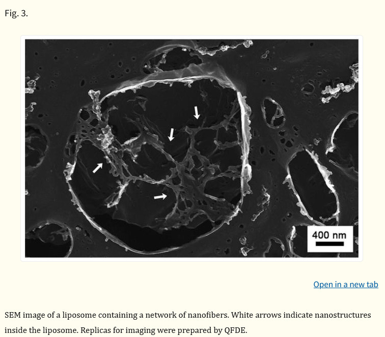
Light-induced self-assembly of nanofibers inside liposomes†
The
liposome is a self-assembled structure where a lipid bilayer surrounds
an aqueous compartment. With a typical volume on the order of one
thousandth of a cubic micron, this interior compartment has been used to
carry drugs, peptides, proteins and DNA for applications in molecular
biology, pharmaceuticals, and cosmetics.1
Beyond the simple containment of molecules, the confined interior of a
liposome is also an interesting space to explore supramolecular
chemistry. In the literature, supramolecular structures that have been
formed inside liposomes include actin fibers2 and fibril-shaped precipitates of the cancer drug doxorubicin.3
These examples, however, typically use ion injection or pH gradients
that depend on the invasive diffusion of ions through the liposomal
membrane in order to stimulate aggregation of molecules. In
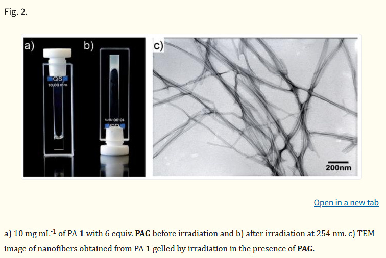
this report, we have investigated the use of light to non-invasively
induce the self-assembly of encapsulated molecules into nanofiber
networks inside liposomes. Bulk nanostructure formation by light has
been observed in the form of photosensitive gels and liquid crystals
Nanowires have been used to create optoelectronics and lasers - as this work by Sandia National Laboratory shows:
1 Non-polar InGaN/GaN core-shell single nanowire lasers
We
report lasing from nonpolar p-i-n InGaN/GaN multi-quantum well
core–shell single-nanowire lasers by optical pumping at room
temperature. The nanowire lasers were fabricated using a hybrid approach
consisting of a top-down two-step etch process followed by a bottom-up
regrowth process, enabling precise geometrical control and high material
gain and optical confinement. The modal gain spectra and the gain
curves of the core–shell nanowire lasers were measured using
micro-photoluminescence and analyzed using the Hakki-Paoli method.
Significantly lower lasing thresholds due to high optical gain were
measured compared to previously reported semipolar InGaN/GaN core–shell
nanowires, despite significantly shorter cavity lengths and reduced
active region volume. Mode simulations show that due to the core–shell
architecture, annular-shaped modes have higher optical confinement than
solid transverse modes. The results show the viability of this p-i-n
nonpolar core–shell nanowire architecture, previously investigated for
next-generation light-emitting diodes, as low-threshold, coherent
UV–visible nanoscale light emitters, and open a route toward monolithic,
integrable, electrically injected single-nanowire lasers operating at
room temperature.
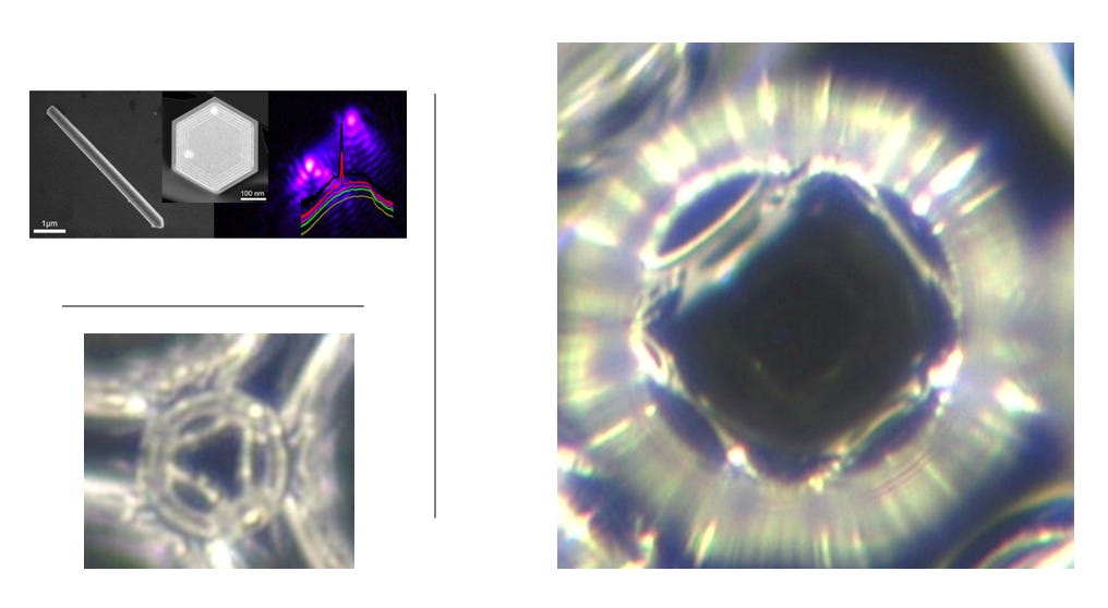
Below
you can see the geometrics of nanowire lasers made from Silicone,
Aluminum, Nickel and other elements. As a comparison are the geometric
shapes that develop within the liposomes. Nanowire lasers are used for
optoelectronics.
Figure
1. Schematic diagram of the InGaN/GaN core-shell nanowire laser
fabrication process, SEM and TEM images of the InGaN/GaN core-shell
nanowire laser. (a) Silica microspheres deposited on the n-type GaN
film. (b) n-type GaN nanowires with tapered and rough sidewalls after
ICP dry etch. (c) Straight and smooth sidewalls of n-type GaN nanowires
are created after AZ400K wet etch. (d) Shell layers (n-GaN layer,
InGaN/GaN MQW, AlGaN electron blocking layer, and p/p+ GaN capping
layer) are grown on the n-type GaN nanowire template. (e) SEM image of a
core-shell nanowire transferred onto a Si3N4/Si substrate.
I have shown the nanofibers/ wires and integrated circuits in this mesogen article.
Summary:
We
are learning more on how liposomes can interact and self assemble
nanowires, biosensors and other nanotechnological devices. Biotechnology
is advancing the pharmaceutical industry in multipurpose scientific
areas. Optoelectronics and biophotonics, plasmonic crystals all are
elements that can be utilized for Artificial Intelligence manipulated
quantum computing, biosensing, genetic engineering and other modalities.
Asking questions as to how these possibilites are affecting humanity is
improtant in order to understand broader implications of how these
nanotechnology substances interact with human physiology - an ultimately
change us on a fundamental unseeen nano and micro level.
No comments:
Post a Comment