MORGELLONS:
FIRST OBSERVATIONS
Clifford E Carnicom
Aug 12 2006
Edited Aug 16 2006
Copyright 2006 by Clifford E Carnicom and Jan Smith
FIRST OBSERVATIONS
Clifford E Carnicom
Aug 12 2006
Edited Aug 16 2006
Copyright 2006 by Clifford E Carnicom and Jan Smith
This paper is being presented in two stages. The
first paper will describe a series of observations under the microscope
at relatively high power. The subject of observation is sample material
received from an individual that displays the symptoms of what is now
known as Morgellons
disease. The Morgellons disease is characterized in
part by the presence of a host of unusual skin conditions, commonly
including persistent lesions and unusual fibers or filaments. The second
paper will be commentary on the Morgellons issue from the perspective
of this researcher; it will be presented separately at a later date.
The illness causes much pain and suffering. The
acceptance of the illness by the formal medical community remains
controversial, despite increasing and widespread evidence of its
existence. This inquiry is prompted by the finding that despite several
years of presumed research on the Morgellons issue, there apparently are
no suitably magnified images of the filaments available to the public.
Despite recent media attention to the issue, it also appears to be
commonly claimed that there are not sufficient resources available to
conduct suitable examinations. This presentation will seek to address
this problem to the degree possible here.

Original Sample Envelope Postmarked Jul 14 2006.
Samples are from the subject directly and were sent to me upon my request.
Materials were received in mid July after lengthy
discussion with the individual. This person has previously made numerous
observations and discussions available to the public; photographs were
limited to a magnification of 200x by the equipment that was available. I
offered to conduct a microscopic study at higher magnification, to
photograph the results and to make this information available to the
public. This is the primary purpose of this first report; no concerted
effort to evaluate the nature of the materials will be made on this
page. This page represents several hours of study under the microscope
of one portion of one sample, and much more work remains to be done. I
may or may not have the means or resources to continue this study and it
is questionable that this work should occur under the domain of citizen
activism.
One of the goals of this paper is to also provide the
reader with a sense of scale, and to show a progression from the
original materials as they exist from the body to the highest
magnification possible with my equipment. The materials received were
packaged carefully, thoroughly labeled and in good order. The subject’s
description of the physical symptoms encountered are thorough and
complete. Any questions put forth to the individual about the illness
have been answered in full candor and detail. The subject has provided
numerous samples to a medical doctor in the past but no specific
response, descriptions, photos or analysis have apparently been provided
in return. Detailed information or replies from any formal medical
representative, non-profit support or research groups, educational
institutions and government agencies appears to be grossly deficient.
There appears to be no adequate response to the individual’s many
appeals for analysis of the illness symptoms and physical
manifestations. The ramifications of a potentially large scale health
issue that may be affecting a much larger portion of the population than
is currently recognized must be considered. In addition to the
suffering that has been endured, those leading individuals that have
come forth with their appeals for assistance have in the main been
derided, denied or refused. At the very least this group requires our
humanitarian compassion and medical assistance.
It is also necessary to confront directly the
numerous claims of “delusion” that are commonly circulated in
conjunction with the public reports on this issue, and to seek out if
there are any agendas that may be associated with this characterization
of the illness. It must also be asked why the citizenry is in the
position of having to provide the information on this page to the
public. The question of unknown health risks to the public (and even to
this researcher) should be addressed.
It is a legitimate question to seek out if these
materials are of any unusual form or nature, as this does not appear to
be properly and publicly addressed at this point. Adequate images under
the microscope may be of help here.
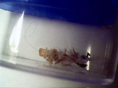
One of several original sample materials received in sealed plastic container.
Materials photographed prior to handling and observation.
Materials stated to come from a lesion on the torso.
Embedded filaments within the lesion material are visible
(isolated fibers just visible to the naked eye).
This container measures approximately 1 inch (2.5cm) across.
Observations to follow are from this sample only.
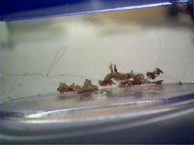
Second photograph of original sample materials received.
Additional samples exist for future analysis.
Several filaments that emanate from the lesion material are visible to the naked eye
Observations to follow are from this sample only.
The fibers that are visible and that emanate from the
lesion material in the photographs above are the subject of the
photographs below. Five magnification levels are available with the
equipment being used: approximately 700x, 1400x, 2800x, 5600x and 8600x.
Digital magnification of the final image can be increased further if
the situation warrants it and if
the image quality supports the enlargement. The limit of conventional
optical magnification is approximately 2000x. The higher levels shown
here have been achieved with the combination of digital camera equipment
(primarily astronomic) and a decent optical microscope. It is believed
that these images are the first available publicly that show internal
detail of the fibers that are apparently representative of the
Morgellons condition. Much deserves to be explained and accounted for
with the disclosures that follow, as it becomes quickly evident that
these are not typical nor uniform fibers.
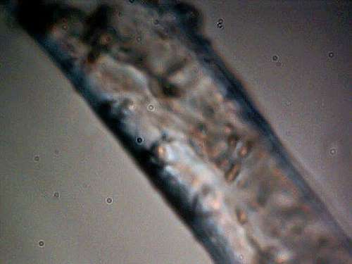
The first image shown below is at a magnification of
approximately 700 times. At the level of 700x, there is relatively
little detail that can be seen. The photograph is adequate, however, to
obtain a first estimate of its width; this first measurement is
approximately 10 to 12 microns. This measurement already seriously
calls into question any claim of these fibers being a human hair, as
they will measure from approximately 60 to 100 microns in width. The
irregular form of the fiber and twisting that is apparent further
eliminates any realistic comparison to a human hair. At this point
recall that most images that have been made available have been at a
level of 200x or less; this already explains why little information
about the appearance of the fibers, let alone any internal structure, is
available to the public to review. I have encountered two extremely high magnification images
taken with an electron microscope, however, it will be seen that no
internal detail is available from those images. As there is no
commentary associated with those images, there can be no further
explanation of that deficiency at this point.
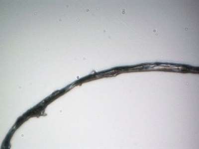
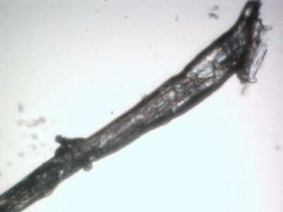
Magnification approximately 700x.
Approximate dimension in width : 10-12 microns.
No major distinguishing characteristics visible.
Indications of some internal structure to fiber may be apparent.
Suitable for measurement and comparison to human hair.
Some unevenness in size noted and ability of the fiber to fold or twist is visible..
Two different fibers examined; both appear essentially identical at this point.
The next presentation will be that of two control
photographs for purposes of comparison and to show the capability of the
modified microscopy equipment that is being used. THESE ARE FOR CONTROL
PURPOSES ONLY AND ARE NOT ASSOCIATED WITH THE SAMPLE MATERIALS IN ANY
WAY. The first photograph will be that of a human hair, also at a
magnification of approximately 700 times. The next photograph will be
that of a human blood cell at approximately 8600 times; a human blood
cell measures on the order of 6-8 microns across. For further
comparison, bacteria are commonly on the order of up to 10 microns in
size, and viruses are usually 1 micron or less. An asbestos fiber is
on the order of 2 microns. For further comparisons and extensive fiber
studies, please consult some of the earlier work on this site.
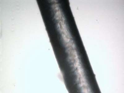
Human Hair : FOR CONTROL PURPOSES ONLY
Magnification approximately 700x..
Note smooth outline and uniform size.
Measurement : approximately 65 microns across.
No significant internal structure or form apparent.
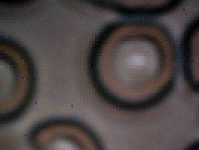
Human Blood Cell : FOR CONTROL PURPOSES ONLY
Magnification approximately 8600x.
Approximate size of cell : 7 microns in diameter
This image represents the upper end of quality
and magnification of the equipment being used in this report.
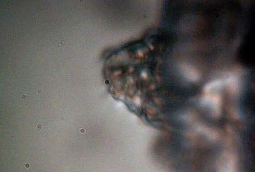
The next set of photographs, the primary focus of
this report, will show a series of photographs at 1400x and 5600x levels
of magnification. At this stage of the research project, I will
largely let the photographs speak for themselves, with minor comments to
assist in their interpretation. The next two photographs at 1400x now
begin to show some interesting features and form that have not been
visible with the initial work. First, it is observed that the fibers
have a much more complicated internal structure than was discernible at
low magnification. In addition, there is more variance in the
dimensions of the fiber than is originally evident. Both of these
factors alone begin to seriously question or eliminate claims of
commonly known fibers, be they artificial or natural. In the media
reports alone, there are now reports of attempted matching of the fiber
form using large forensic databases and a complete failure of
identification in that attempt. One of the objectives of this report is
to allow the public itself to see why that failure is likely occurring.
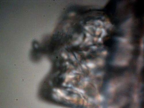
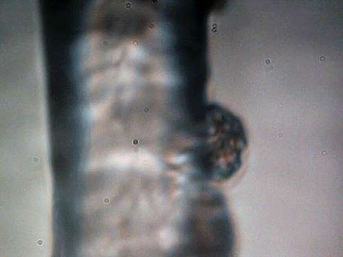
There is a second revelation at this level of
magnification and observation. What appears to be a single filament
coming from the lesion material is actually much more complicated and
that much exists that is not visible to the naked eye. In the second of
these two photographs, notice the rather complicated web of numerous
fibers. This arrangement was most certainly not visible by eye when
this sample was placed under the microscope. It is at this point that
much greater interest is to be attached to the identification of these
filaments, as well as whatever structures may be contained within. We
also notice, particularly in the second photograph of greater
translucency, that internal, much smaller structures of elliptical form
exist. This begins to strongly suggest a biological nature to the
fibers, and a case for ruling out human hairs as well as any common
fiber form, natural or artificial, is now made.
At this point, it is at least appropriate to address
the rather massive efforts that have been and that are being made to
characterize this illness as a psychological problem of the afflicted
individuals. This effort will be addressed more completely in the
commentary section that shall follow at a later time. In the interim,
however, if the materials being shown here are representative of the
Morgellons condition, such efforts to foist a perception of “delusion”
upon the public can only be interpreted as a ruse of the highest order
in an effort to conceal, deny and avoid the true issues that we are
facing. The reports of occurrence of this illness are increasing
and they are global at this stage. It is reasonable to inquire as to
what agendas may be in place to so forcefully attempt to influence the
public perception of this condition or disease.
Magnification approximately 1400x.
Note variation in fiber form and internal structure becoming evident.
Notice irregularities on the surface of the fiber.
Notice translucent quality of the fiber.
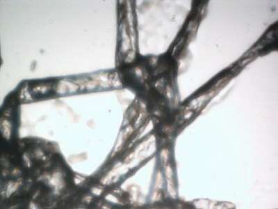
Magnification approximately 1400x.
Numerous fibers are now available; this conglomerate not visible to the naked eye.
Notice internal structures becoming increasingly visible.
Biological natures are more strongly indicated at this point of observation.
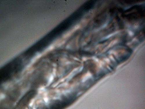
The next and final set of photographs will be at
5600x. Several important discoveries take place. It is now quite
common within certain segments of the primary fiber to find an internal
sub-fibrous structure. It can now be seen that what appears to be a
single fiber is composed of innumerable sub-fibers, and that these
sub-fibers measure at the micron or sub-micron level. There is no known
previous disclosure of this fact on the Morgellons condition and a much
more complex interpretation of the actual nature of the fibers must now
be proposed. Secondly, internal spherical or elliptical structures now
appear within the primary fiber, measuring on the order of 1 micron
(virus size). It is now a compelling priority to identify these
structures and their functions, including the internal micron sized
sub-fibers.
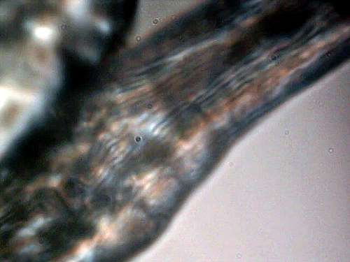
Magnification approximately 5600x.
Notice internal filament structure within the fiber.
Width of the internal fibrous structure is at the micron or sub-micron level.
Magnification approximately 5600x.
Notice internal generally circular structures.
Strongly indicative of a biological nature at this point.
These structures measure on the order of 1 micron (virus size).
Increasingly complex internal nature of the original sample fiber is now evident.
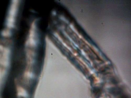
The last major discovery by observation at this point
is what appears to a “budding” structure of some sort. These
structures appear on the edge of the fiber at irregular intervals.
These structures contain two further components within. The first of
these are spherical or elliptical structures at the micron level within
an encasing, translucent shell. In addition, innumerable fibers at the
sub-micron level emerge from the budding structure. The budding
structures are highly indicative of a growth or reproductive process,
and they may be related to the spread of the disease.
Magnification approximately 5600x.
“Budding” structures are apparent on the sides of the fiber at occasional locations.
The budding structures contain internal structures at the roughly micron or sub-micron size.
Budding structures also often contain innumerable filaments within,
measuring apparently at the sub-micron level (Limit of equipment reached).
Reproduction and growth of the primary fiber structure may be closely linked to these budding structures.
The budding structures generally appear to be quite complex in form, structure and organization.
Magnification approximately 5600x.
Complex internal organization of sub-fibers and structural forms is apparent.
Magnification approximately 5600x.
This photograph shows the ability of the fiber to be folded and/or twisted.
Internal parallel organization of sub-fibers is visible.
Non-uniformity of the fibers dimensions is also evident.
Transverse separation or structure also visible in lower right of image.
Magnification approximately 5600x.
Additional budding structure visible on the edge of the primary fiber.
Complex internal micron size structures within.
Translucent encasement that is indicative or suggestive of reproductive capability.
Magnification approximately 5600x.
Additional budding structure visible on the edge of the primary fiber.
Complex internal micron size structures within.
The conclusion of this report is necessarily brief at
this time. The basic conclusions that can be made are as follows.
First, there has been a complete failure of the formal medical
community, non-profit organizations and government to adequately
research and distribute information to the public on the nature of the
Morgellons condition. If the samples studied and shown here are in any
way representative of the Morgellons disease, they show that any effort
to influence the public to accept this evidence as being of
psychological origin or as insignificant are disingenuous at the
highest level. Any motive of secrecy and or misinformation is to be
confronted directly and disclosed. The so-called efforts at research by
various organizations, including non-profit, university and government
are to be called into question; there is a serious lack of informing the
public as to the basic nature of the condition. No citizen should be
assuming the risk of attempting to identify the nature of this illness.
The traditional medical community and government health organizations
have already displayed an appalling failure of addressing the urgency of
this matter. I call upon all of those individuals or groups with the
proper resources to strike to the core of this issue as quickly as
possible, and to disclose all results of the findings to the public as
they occur.
Clifford E Carnicom
August 12 2006
August 12 2006
Notes:
Additional research and or information from other sources will be linked into this report as it becomes available.
Additional commentary of the general state of findings on the Morgellons issue will be presented at a later date.
The results of this report are of a preliminary nature, and they are restricted to the materials that have been provided by a single individual.

No comments:
Post a Comment